
User:SchwarzeSchlange/Personal sandbox
Let's try it all!
| Nucleus accumbens | |
|---|---|
| Details | |
| Identifiers | |
| Latin | nucleus accumbens septi |
| Anatomical terms of neuroanatomy | |
The nucleus accumbens (NAcc), formerly known as the nucleus accumbens septi (Latin for nucleus leaning against the septum), is a bilateral structure of the basal forebrain, consisting of a mass of neurons in the ventral part of the striatum [1] It is known to play an important role in reward and addiction,[2][3][4] and in the placebo effect [5] (although an involvement of NAcc in aversive learning such as fear learning [6] and taste aversion learning [7] has been also documented).
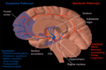
It is located underneath the anterior limb of the internal capsule, lateral to the vertical part of the diagonal band of Broca, and medial to the claustrum and piriform cortex. The NAcc extends dorsolaterally into the ventral putamen and dorsomedially into the ventral caudate without clear boundaries.[8] Together with the olfactory tubercle, the NAcc forms the ventral striatum, which is part of the basal ganglia.[9]
The nucleus accumbens can be divided into two structures—the nucleus accumbens core and the nucleus accumbens shell. These structures have different morphology and function.
Divisions[edit]
According mainly to hodological and immunohistochemical considerations, the NAcc can be divided into a shell and a core subregions.
NAcc shell[edit]
Immunoreactivity to calbidin-D28k has been regarded as a reliable marker to distinguish the NAcc subterritories.[1] Thus, the medial, ventral and lateral parts of the NAcc, which are lightly to moderately immunoreactive to calbindin-D28k, form the NAcc shell.[1][10] However, a weaker (compared to the core) immunoreactivity for dopamine receptor D2[1] (but see [11]) in the NAcc shell has been also found. On the contrary, a stronger immunoreactivity (compared to the core) for substance P, dopamine receptor D1 and D3, enkephalin, and neurotensin is characteristic of the NAcc shell.[1]
Cell types[edit]
The principal neuronal cell type found in the nucleus accumbens, which make up approximately 90% of all accumbal neurons, is the medium spiny neuron.[12] Except for a slight decrease in diameter (8-20 μm for accumbal to 12-18 μm in the striatum), no signficant differences exist between accumbal and striatal medium spiny neurons.[12]
While 95% of the neurons in the nucleus accumbens are medium spiny GABA-ergic projection neurons, other neuronal types are also found such as large aspiny cholinergic interneurons.
Output and input[edit]
Output[edit]
The output neurons of the nucleus accumbens send axon projections to the ventral analog of the globus pallidus, known as the ventral pallidum (VP). The VP, in turn, projects to the medial dorsal nucleus of the dorsal thalamus, which projects to the prefrontal cortex as well as the striatum. Other efferents from the nucleus accumbens include connections with the substantia nigra and the pontine reticular formation.
Input[edit]
Major inputs to the nucleus accumbens include prefrontal association cortices, basolateral amygdala, and dopaminergic neurons located in the ventral tegmental area (VTA), which connect via the mesolimbic pathway. Thus the nucleus accumbens is often described as one part of a cortico-striato-thalamo-cortical loop.
Dopaminergic input from the VTA is thought to modulate the activity of neurons within the nucleus accumbens. These terminals are also the site of action of highly-addictive drugs such as cocaine and amphetamine, which cause a manifold increase in dopamine levels in the nucleus accumbens. In addition to cocaine and amphetamine, almost every recreational drug has been shown to increase dopamine levels in the nucleus accumbens.
Another major source of input comes from the CA1 and ventral subiculum of the hippocampus to the dorsomedial area of the Nucleus accumbens. The neurons of the hippocampus have a noteworthy correlation to slight depolarizations of cells in the nucleus accumbens, which makes them more positive and therefore more excitable. The correlated cells of these excited states of the medium spiny neurons in the Nucleus accumbens are shared equally between the subiculum and CA1. The subiculum neurons are found to hyperpolarize(increase negativity) while the CA1 neurons "ripple"(fire > 50 Hz) in order to accomplish this priming.[13]
Research[edit]
In the 1950s, James Olds and Peter Milner implanted electrodes into the septal area of the rat and found that the rat chose to press a lever which stimulated it. It continued to prefer this even over stopping to eat or drink. This suggests that the area is the 'pleasure center' of the brain. The septal nuclei are not directly connected to the nucleus accumbens, however.[14]
Although the nucleus accumbens has traditionally been studied for its role in addiction, it plays an equal role in processing many rewards such as food and sex. Interestingly, the nucleus accumbens is selectively activated during the perception of pleasant, emotionally arousing pictures and during mental imagery of pleasant, emotional scenes.[15][16] A recent study found that it is involved in the regulation of emotions induced by music,[17] perhaps consequent to its role in mediating dopamine release. The nucleus accumbens plays a role in rhythmic timing and is considered to be of central importance to the limbic-motor interface (Mogensen).
In April 2007, two research teams reported on having inserted electrodes into the nucleus accumbens in order to use deep brain stimulation to treat severe depression.[18]
In addition, in July 2007, researcher Jon-Kar Zubieta published findings that the nucleus accumbens is central to the machinery of the placebo effect. His group has confirmed that specific neural circuits and neurotransmitter systems respond to the expectation of benefit during placebo administration and that these expectations induce measurable physiological changes.[19]
The nucleus accumbens has been targeted by stereotactic surgery for ablation as a treatment in China for alcoholism.[20]
Additional images[edit]
-
 Dopamine and serotonin
 -
 MRI coronal slice showing nucleus accumbens outlined in red


References[edit]
- ^ a b c d e
Groenewegen, H.J., Wright, C.I., Beijer, A.V.J., Voorn, P. (1999). Ann. N. Y. Acad. Sci. 877: 49–63. doi:10.1111/j.1749-6632.1999.tb09260.x. PMID 10415642.
{{cite journal}}: Missing or empty|title=(help)CS1 maint: multiple names: authors list (link) - ^ Di Chiara, G. (2002). "Nucleus accumbens shell and core dopamine: differential role in behavior and addiction". Behav. Brain Res. 137 (1–2): 75–114. doi:10.1016/S0166-4328(02)00286-3. PMID 12445717.
- ^ Kelley, A.E. (2004). "Ventral striatal control of appetitive motivation: role in ingestive behavior and reward-related learning". Neurosci. Biobehav. Rev. 27 (8): 765–776. doi:10.1016/j.neubiorev.2003.11.015. PMID 15019426.
- ^ Carelli, R.M. (2004). "Nucleus accumbens cell firing and rapid dopamine signaling during goal-directed behaviors in rats". Neuropharmacology. 47 (Suppl. 1): 180–189. doi:10.1016/j.neuropharm.2004.07.017. PMID 15464136.
- ^
Scott, D.J., Stohler CS, Egnatuk, C.M., Wang, H., Koeppe, R.A., Zubieta, J.K. (2007). "Individual differences in reward responding explain placebo-induced expectations and effects". Neuron. 55 (2): 325–336. doi:10.1016/j.neuron.2007.06.028. PMID 17640532.
{{cite journal}}: CS1 maint: multiple names: authors list (link) - ^
- Schwienbacher, I., Fendt, M., Richardson, R., Schnitzler, H.U. (2004). "Temporary inactivation of the nucleus accumbens disrupts acquisition and expression of fear-potentiated startle in rats". Brain Res. 1027 (1–2): 87–93. doi:10.1016/j.brainres.2004.08.037. PMID 15494160.
{{cite journal}}: CS1 maint: multiple names: authors list (link) - Riedel, G., Harrington, N.R., Hall, G., Macphail, E.M. (1997). "Nucleus accumbens lesions impair context, but not cue, conditioning in rats". Neuroreport. 8 (11): 2477–2481. PMID 9261812.
{{cite journal}}: CS1 maint: multiple names: authors list (link) - Haralambous, T., Westbrook, R.F. (1999). "An infusion of bupivacaine into the nucleus accumbens disrupts the acquisition but not the expression of contextual fear conditioning". Behav. Neurosci. 113 (5): 925–940. PMID 10571476.
{{cite journal}}: CS1 maint: multiple names: authors list (link) - Ammassari-Teule, M., Passino, E., Restivo, L., de Marsanich, B. (2000). "Fear conditioning in C57/BL/6 and DBA/2 mice: variability in nucleus accumbens function according to the strain predisposition to show contextual- or cue-based responding". Eur. J. Neurosci. 12 (12): 4467–4474. doi:10.1111/j.1460-9568.2000.01333.x. PMID 11122357.
{{cite journal}}: CS1 maint: multiple names: authors list (link) - Jongen-Rêlo, A.L., Kaufmann, S., Feldon, J. (2003). "A differential involvement of the shell and core subterritories of the nucleus accumbens of rats in memory processes". Behav. Neurosci. 117 (1): 150–168. PMID 12619918.
{{cite journal}}: CS1 maint: multiple names: authors list (link)
- Schwienbacher, I., Fendt, M., Richardson, R., Schnitzler, H.U. (2004). "Temporary inactivation of the nucleus accumbens disrupts acquisition and expression of fear-potentiated startle in rats". Brain Res. 1027 (1–2): 87–93. doi:10.1016/j.brainres.2004.08.037. PMID 15494160.
- ^
- Mark, G.P., Blander, D.S., Hoebel, B.G.. (1991). "A conditioned stimulus decreases extracellular dopamine in the nucleus accumbens after the development of a learned taste aversion". Brain Res. 551 (1–2): 308–310. doi:10.1016/0006-8993(91)90946-S. PMID 1913157.
{{cite journal}}: CS1 maint: multiple names: authors list (link) - Ramírez-Lugo, L., Zavala-Vega, S., Bermúdez-Rattoni, F. (2006). "NMDA and muscarinic receptors of the nucleus accumbens have differential effects on taste memory formation". Learn. Mem. 13 (1): 45–51. doi:10.1101/lm.103206. PMID 16452653.
{{cite journal}}: CS1 maint: multiple names: authors list (link) - Pedroza-Llinás, R., Ramírez-Lugo, L., Guzmán-Ramos, K., Zavala-Vega, S., Bermúdez-Rattoni, F. (2009). "Safe taste memory consolidation is disrupted by a protein synthesis inhibitor in the nucleus accumbens shell". Neurobiol. Learn. Mem. 92 (1): 45–52. doi:10.1016/j.nlm.2009.02.011. PMID 19249379.
{{cite journal}}: CS1 maint: multiple names: authors list (link)
- Mark, G.P., Blander, D.S., Hoebel, B.G.. (1991). "A conditioned stimulus decreases extracellular dopamine in the nucleus accumbens after the development of a learned taste aversion". Brain Res. 551 (1–2): 308–310. doi:10.1016/0006-8993(91)90946-S. PMID 1913157.
- ^ Neto, L.L., Oliveira, E., Correia, F., Ferreira, A.G. (2008). "The human nucleus accumbens: where is it? A stereotactic, anatomical and magnetic resonance imaging study". Neuromodulation: Technology at the Neural Interface. 11 (1): 13–22. doi:10.1111/j.1525-1403.2007.00138.x.
{{cite journal}}: CS1 maint: multiple names: authors list (link) - ^ Nucleus Accumbens
- ^
Prensa, L., Richard, S., Parent, A. (2003). "Chemical anatomy of the human ventral striatum and adjacent basal forebrain structures". J. Comp. Neurol. 460 (3): 345–367. doi:10.1002/cne.10627. PMID 12692854.
{{cite journal}}: CS1 maint: multiple names: authors list (link) - ^ Pickel, V.M., Chan, J., Kearn, C.S., Mackie, K. (2006). "Targeting dopamine D2 and cannabinoid-1 (CB1) receptors in rat nucleus accumbens". J. Comp. Neurol. 495 (3): 299–313. doi:10.1002/cne.20881. PMID 16440297.
{{cite journal}}: CS1 maint: multiple names: authors list (link) - ^ a b Meredith, C.E. (1999). "The synaptic framework for chemical signaling in nucleus accumbens". Ann. N. Y. Acad. Sci. 877: 140–156. doi:10.1111/j.1749-6632.1999.tb09266.x. PMID 10415648.
- ^ O'Donnell, P., Goto, Y. (2001). "Synchronous activity in the hippocampus and nucleus accumbens in vivo". J. Neurosci. 21 (4): RC131. PMID 11160416.
{{cite journal}}: CS1 maint: multiple names: authors list (link) - ^ Olds J, Milner P (1954). "Positive reinforcement produced by electrical stimulation of septal area and other regions of rat brain". J Comp Physiol Psychol. 47 (6): 419–27. doi:10.1037/h0058775. PMID 13233369. article
- ^ Costa, VD, Lang, PJ, Sabatinelli, D, Bradley MM, and Versace, F (2010). "Emotional imagery: Assessing pleasure and arousal in the brain's reward circuitry". Human Brain Mapping. 31 (9): 1446–1457. doi:10.1002/hbm.20948. PMID 20127869.
{{cite journal}}: CS1 maint: multiple names: authors list (link) - ^ Sabatinelli, D, Lang, PJ, Bradley, MM, Costa, VD, and Versace, F (2007). "Pleasure rather than salience activates human nucleus accumbens and medial prefrontal cortex". Journal of Neurophysiology. 98 (9): 1374–1379. PMID 17596422.
{{cite journal}}: Text "doi:10.1152/jn.00230.2007" ignored (help)CS1 maint: multiple names: authors list (link) - ^ Menon, Vinod & Levitin, Daniel J. (2005) The rewards of music listening: Response and physiological connectivity of themesolimbic system." NeuroImage 28(1), pp. 175-184
- ^ Brain Electrodes Help Treat Depression, Technology Review, 26 April 2007
- ^ http://www.eurekalert.org/pub_releases/2007-07/cp-brc071607.php Brain region central to placebo effect identified
- ^ Wu HM, Wang XL, Chang CW, Li N, Gao L, Geng N, Ma JH, Zhao W, Gao GD. (2010). Preliminary findings in ablating the nucleus accumbens using stereotactic surgery for alleviating psychological dependence on alcohol. Neurosci Lett. 473: 77–81 doi:10.1016/j.neulet.2010.02.019 PMID 20156524
External links[edit]
- The role of the nucleus accumbens in the reward circuit. Part of "The Brain From Top to Bottom." at thebrain.mcgill.ca
- Nucleus Accumbens - Cell Centered Database
- Stained brain slice images which include the "nucleus%20accumbens" at the BrainMaps project