
File:Fmicb-08-01337-g001.jpg

Size of this preview: 800 × 590 pixels. Other resolutions: 320 × 236 pixels | 640 × 472 pixels | 1,024 × 755 pixels | 1,280 × 944 pixels | 1,800 × 1,328 pixels.
Original file (1,800 × 1,328 pixels, file size: 221 KB, MIME type: image/jpeg)
| This is a file from the Wikimedia Commons. Information from its description page there is shown below. Commons is a freely licensed media file repository. You can help. |
Summary
| Description |
English: Schematic representation of baculovirus structure and infection cycle. (A) Nucleopolyhedrovirus occlusion bodies (OBs) are polyhedral proteinaceous bodies, mainly comprised of crystalline polyhedrin that surrounds occlusion derived virions (ODVs). The ODVs contain either a single nucleocapsid (single type) or between one and several nucleocapsids (multiple type) in each ODV. For granuloviruses the OB is granule-shaped and contains a single ODV with a single nucleocapsid surrounded by the crystalline protein granulin. In all cases each nucleocapsid contains a single viral genome. (B) Sequential steps of nucleopolyhedrovirus transmission and replication. During primary infection, (A) OBs are ingested during feeding on contaminated foliage. (B) OBs are solubilized in the insect midgut and release ODVs that pass through the peritrophic membrane (C) and fuse with the microvilli of midgut epithelial cells (D). Nucleocapsids travel to the nucleus where they release the viral genome to initiate replication. (E) Virus replication occurs in virogenic stroma. Progeny nucleocapsids assemble and bud through the basal membrane (F) during which they acquire an envelope containing GP64 or F fusion protein present in the virus-modified cell membrane. During the secondary phase of infection these budded virions (BVs) disperse in the hemolymph or along the cells of the insect traqueal system (traqueoblasts) to spread the infection to the cells of other tissues in the insect. (G) BVs enter cells by endocytosis and replicate in the nucleus. Newly assembled nucleocapsids (H) may bud out of the cell or may be enveloped to form ODVs that are occluded into OBs (I). At the end of the infectious cycle OBs accumulate in the nucleus (J). Upon death the larvae typically hang from the uppermost leaves of the host plant (K), the larval tegument ruptures and releases OBs that contaminate foliage for further cycles of horizontal transmission. |
| Date | |
| Source | https://www.frontiersin.org/articles/10.3389/fmicb.2017.01337/full |
| Author | Trevor Williams, Cristina Virto, Rosa Murillo, and Primitivo Caballero |
Licensing
This file is licensed under the Creative Commons Attribution 4.0 International license.
- You are free:
- to share – to copy, distribute and transmit the work
- to remix – to adapt the work
- Under the following conditions:
- attribution – You must give appropriate credit, provide a link to the license, and indicate if changes were made. You may do so in any reasonable manner, but not in any way that suggests the licensor endorses you or your use.
File history
Click on a date/time to view the file as it appeared at that time.
| Date/Time | Thumbnail | Dimensions | User | Comment | |
|---|---|---|---|---|---|
| current | 06:01, 26 June 2020 | 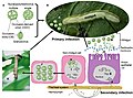 | 1,800 × 1,328 (221 KB) | Guest2625 | Uploaded a work by Trevor Williams, Cristina Virto, Rosa Murillo, and Primitivo Caballero from https://www.frontiersin.org/articles/10.3389/fmicb.2017.01337/full with UploadWizard |
File usage
The following pages on the English Wikipedia use this file (pages on other projects are not listed):
Global file usage
The following other wikis use this file:
- Usage on tr.wikipedia.org