
File:Computed tomography of human brain - large.png

Original file (3,639 × 2,595 pixels, file size: 3.9 MB, MIME type: image/png)
| This is a file from the Wikimedia Commons. Information from its description page there is shown below. Commons is a freely licensed media file repository. You can help. |
| This is a featured picture, which means that members of the community have identified it as one of the finest images on the English Wikipedia, adding significantly to its accompanying article. If you have a different image of similar quality, be sure to upload it using the proper free license tag, add it to a relevant article, and nominate it. |
| This image was selected as picture of the day on the English Wikipedia for July 11, 2008. |
| This file is made available under the Creative Commons CC0 1.0 Universal Public Domain Dedication. | |
| The person who associated a work with this deed has dedicated the work to the public domain by waiving all of their rights to the work worldwide under copyright law, including all related and neighboring rights, to the extent allowed by law. You can copy, modify, distribute and perform the work, even for commercial purposes, all without asking permission.
|
|

| Description |
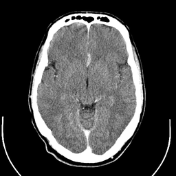
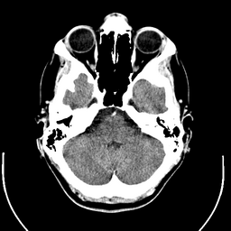
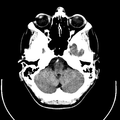
English: Computer tomography of human brain, from base of the skull to top. Taken with intravenous contrast medium.
It was taken Mars 23, 2007 on the uploader, after a 20 minute episode of homonymous hemianopsia with loss of the left visual field, but nothing strange was found. Three episodes of scotoma occurred in the following years, whereof the last one was scintillating (depiction). Otherwise, there were no further neurological symptoms.
Türkçe: Geçirdiği bir kaza neticesinde homonim hemianopsi vakası oluşan bir hastanın beyninin bilgisayarlı tomografisi. Tomografi neticesinde bir anomaliye rastlanmamıştır. |
| Date | Uploaded January 17, 2008 |
| Source | Radiology, Uppsala University Hospital. Uploaded by Mikael Häggström. |
| Author | Department of Radiology, Uppsala University Hospital. Uploaded by Mikael Häggström. |
| Permission (Reusing this file) |
Compound images
-

-
 Inverted
Scrollable stack
For larger version, see Category:Computed tomography images of Mikael Häggström's brain. To move through the images, hover over the image and use scroll wheel, drag the mouse, or click the < or the > above each stack. This functionality should activate when the page is fully loaded, which may take some time.


Case with multiplanar reconstruction
-
 Brain, case 1: Multiplanar, but no intravenous contrast.
Individual images


Licencing
| This file is made available under the Creative Commons CC0 1.0 Universal Public Domain Dedication. | |
| The person who associated a work with this deed has dedicated the work to the public domain by waiving all of their rights to the work worldwide under copyright law, including all related and neighboring rights, to the extent allowed by law. You can copy, modify, distribute and perform the work, even for commercial purposes, all without asking permission.
|
DICOM format
File history
Click on a date/time to view the file as it appeared at that time.
| Date/Time | Thumbnail | Dimensions | User | Comment | |
|---|---|---|---|---|---|
| current | 01:11, 24 December 2017 |  | 3,639 × 2,595 (3.9 MB) | Shashi. | Reverted to version as of 12:49, 1 February 2008 (UTC) |
| 10:59, 8 May 2008 | 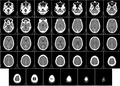 | 3,639 × 2,595 (3.17 MB) | CountingPine | Optimise using PNGOUT | |
| 12:49, 1 February 2008 |  | 3,639 × 2,595 (3.9 MB) | Mikael Häggström | {{34 computer tomography images}} {{Individual images of CT of Mikael Häggström's brain}} | |
| 11:56, 31 January 2008 |  | 3,639 × 2,595 (4.03 MB) | Mikael Häggström | {{34 computer tomography images}} {{Individual images of CT of Mikael Häggström's brain}} |
File usage
- CT scan
- Computed tomography of the head
- Reconstruction from projections
- User:Fitness queen04/sandbox
- User:Flyer22 Frozen/Human brain
- User:Mikael Häggström
- User:Mikael Häggström/Gallery
- User:VGrigas (WMF)/Quality Media
- User:Wouterstomp/test
- User talk:Mikael Häggström/Archive 1
- User talk:Rhododendrites/Reconsidering FPC on the English Wikipedia
- Wikipedia:Featured picture candidates/CT of brain of Mikael Häggström.png
- Wikipedia:Featured picture candidates/February-2008
- Wikipedia:Featured pictures/Sciences/Biology
- Wikipedia:Featured pictures thumbs/10
- Wikipedia:Picture of the day/July 2008
- Wikipedia:WikiProject Anatomy/Recognized content
- Wikipedia:WikiProject Anatomy/Resources
- Wikipedia:WikiProject Medicine/Recognized content
- Wikipedia:WikiProject Neuroscience
- Wikipedia:Wikipedia Signpost/2008-02-11/Features and admins
- Wikipedia:Wikipedia Signpost/2008-02-11/SPV
- Wikipedia:Wikipedia Signpost/2013-10-02/Op-ed
- Wikipedia:Wikipedia Signpost/Single/2013-10-02
- Wikipedia talk:WikiProject Anatomy/Archive 9
- Template:POTD/2008-07-11
- Portal:Medicine
- Portal:Medicine/Recognized content
- Portal:Medicine/Selected picture
- Portal:Medicine/Selected picture/9
- Portal:Medicine/Selected picture/9, 2008
- Portal:Medicine/Selected picture archive
Global file usage
The following other wikis use this file:
- Usage on bn.wikipedia.org
- Usage on bo.wikipedia.org
- Usage on ca.wikipedia.org
- Usage on es.wikipedia.org
- Usage on fi.wikipedia.org
- Usage on he.wikipedia.org
- Usage on hy.wikipedia.org
- Usage on hyw.wikipedia.org
- Usage on id.wikipedia.org
- Usage on is.wikipedia.org
- Usage on ja.wikipedia.org
- Usage on ka.wikipedia.org
- Usage on kk.wikipedia.org
- Usage on ks.wikipedia.org
- Usage on la.wikipedia.org
- Usage on lt.wikipedia.org
- Usage on pl.wikipedia.org
- Usage on pt.wikipedia.org
- Usage on ro.wikipedia.org
- Usage on ru.wikipedia.org
- Usage on sl.wikipedia.org
- Usage on tr.wikipedia.org
- Bilgisayarlı tomografi
- Vikipedi:Seçkin resimler/Bilim/Biyoloji
- Vikipedi:Seçkin resim adayları/Computed tomography of human brain - large.png
- Vikipedi:Seçkin resim adayları/Arşiv/Ağustos 2008
- Vikipedi:Günün seçkin resmi/Mart 2009
- Vikipedi:Seçkin resimler/Ana sayfaya çıkmış resimler/2009 listesi
- Şablon:GSR/2009-03-20
- Vikipedi:Günün seçkin resmi/Mayıs 2010
- Şablon:GSR/2010-05-15
- Vikipedi:Seçkin resimler/Ana sayfaya çıkmış resimler/2010 listesi
- Vikipedi:Günün seçkin resmi/Ağustos 2018
- Şablon:GSR/2018-08-08
- Usage on uz.wikipedia.org
- Usage on vi.wikipedia.org
View more global usage of this file.