
File:Catopsbaatar skull diagrams.jpg

Original file (5,744 × 1,731 pixels, file size: 3.67 MB, MIME type: image/jpeg)
| This is a file from the Wikimedia Commons. Information from its description page there is shown below. Commons is a freely licensed media file repository. You can help. |
Summary
| Description |
Left (originally fig. 6.): Catopsbaatar catopsaloides, reconstruction of the skull, based on all available specimens, in dorsal view. The nasal vascular foramina are shown as preserved in PM 120/107. In other specimens there may be only two pairs of nasal vascular foramina. Middle (originally fig. 8.): Catopsbaatar catopsaloides, reconstruction of the skull, based on all available specimens, in ventral view. The reconstruction is of an adult individual, but the upper premolars P1 and P3, which disappear during ontogeny, have been reconstructed. The teeth in maxilla are P1, P3, P4, M1, and M2. The choanal region and the middle part of the basicranial region (diagrammatical), including recognition of most of the foramina not preserved or poorly preserved in Catopsbaatar (see text for details), have been reconstructed on the basis of other djadochtatherioid genera, especially Kamptobaatar (Kielan−Jaworowska 1971), Nemegtbaatar (Kielan−Jaworowska et al. 1976), and Kryptobaatar (Wible and Rougier 2000). Right (originally fig. 7.): Catopsbaatar catopsaloides, reconstruction of the skull, based on all available specimens, in lateral view. The reconstruction is of an adult individual, but the upper premolars P1 and P3, which disappear during ontogeny, are reconstructed. The bones of the orbit, not preserved in ZPAL and PM specimens and very fragmentarily preserved only in PIN 4537/4 and /5, have not been reconstructed. The dorsal part of the maxilla/squamosal suture is tentatively reconstructed on the basis of PIN 4537/5 (Fig. 3C, D) and a comparison with Djadochtatherium (Fig. 11B). The arrow points to the posterior zygomatic “ridge” (muscle scar), preserved on the squamosal above the glenoid fossa and discernible in occipital view. The teeth not marked on the dentary are p4 and m1, on maxilla P4 and M1. |
| Date | |
| Source | Kielan−Jaworowska, Z., Hurum, J.H., and Lopatin, A.V. 2005. Skull structure in Catopsbaatar and the zygomatic ridges in multituberculate mammals. Acta Palaeontologica Polonica 50 (3): 487–512. |
| Author | Bogusław Waks−mundzki, pubklished by Kielan−Jaworowska, Z., Hurum, J.H., and Lopatin, A.V. |
Licensing
- You are free:
- to share – to copy, distribute and transmit the work
- to remix – to adapt the work
- Under the following conditions:
- attribution – You must give appropriate credit, provide a link to the license, and indicate if changes were made. You may do so in any reasonable manner, but not in any way that suggests the licensor endorses you or your use.
- share alike – If you remix, transform, or build upon the material, you must distribute your contributions under the same or compatible license as the original.
File history
Click on a date/time to view the file as it appeared at that time.
| Date/Time | Thumbnail | Dimensions | User | Comment | |
|---|---|---|---|---|---|
| current | 01:19, 3 December 2019 | 5,744 × 1,731 (3.67 MB) | FunkMonk | Perhaps a better arrangement. | |
| 22:36, 4 March 2018 | 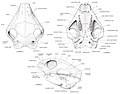 | 3,809 × 2,990 (3.77 MB) | FunkMonk | =={{int:filedesc}}== {{Information |description=Upper left (originally fig. 6.): Catopsbaatar catopsaloides, reconstruction of the skull, based on all available specimens, in dorsal view. The nasal vascular foramina are shown as preserved in PM 120/107. In other specimens there may be only two pairs of nasal vascular foramina. Upper right (originally ig. 8.): Catopsbaatar catopsaloides, reconstruction of the skull, based on all available specimens, in ventral view. The reconstruction is of an adult individ− ual, but the upper premolars P1 and P3, which disappear during ontogeny, have been reconstructed. The teeth in maxilla are P1, P3, P4, M1, and M2. The choanal region and the middle part of the basicranial region (diagrammatical), including recognition of most of the foramina not preserved or poorly pre− served in Catopsbaatar (see text for details), have been reconstructed on the basis of other djadochtatherioid genera, especially Kamptobaatar (Kielan−Jaworowska 1971), Nemegtbaa... |
File usage
Global file usage
The following other wikis use this file:
- Usage on pl.wikipedia.org