
File:Braincases of Baryonyx and Ceratosuchops.jpg

Size of this preview: 450 × 599 pixels. Other resolutions: 180 × 240 pixels | 360 × 480 pixels | 577 × 768 pixels | 769 × 1,024 pixels | 1,538 × 2,048 pixels | 2,761 × 3,677 pixels.
Original file (2,761 × 3,677 pixels, file size: 2.93 MB, MIME type: image/jpeg)
| This is a file from the Wikimedia Commons. Information from its description page there is shown below. Commons is a freely licensed media file repository. You can help. |
Summary
| Description | Braincases of (a, c, e, f, h) Baryonyx walkeri (NHMUK PV R9951) and (b, d, g, i) Ceratosuchops inferodios (IWCMS 2014.95.1–3), in (a, b) right anterolateral and (c, d) right posterolateral, (e–g) right lateral and (h, i) posterior (right side) views, showing the major neurovascular features (and associated foramina) and braincase anatomy. Note that in Baryonyx, the separate cranial nerve trunks X—XI and XII open into a common fossa lateral to the occipital condyle, which is depicted here. Abbreviations: ce, cranial endocast; cul, cultriform process; ct, crista tuberalis; BO, basioccipital; BS, basisphenoid; car, cerebral internal carotid artery canal; F, frontal; fm, foramen magnum; fo, fenestra ovalis; lab, endosseous labyrinth; LS, laterosphenoid; ocv, orbitocerebral vein; oc, occipital condyle; OS, orbitosphenoid; OT, otoccipital; otc, olfactory tract; P, parietal; pit, pituitary; pmcv, posterior middle cerebral vein canal; PRO, prootic; II, optic nerve canal; IV, trochlear nerve canal; V, trigeminal nerve canal; V1, ophthalmic nerve canal; VI, abducens nerve canal; VII, facial nerve canal; IX, glossopharyngeal nerve canal; X–XI, shared canal for the vagus and accessory nerves, and accompanying vessels; XII, hypoglossal nerve canal; ?, potential accessory hypoglossal nerve or venous canal. Scale bars: (a–d) 20 mm and (e–i) 50 mm. |
| Date | |
| Source | https://onlinelibrary.wiley.com/doi/10.1111/joa.13837 |
| Author | Chris Tijani Barker, Darren Naish, Jacob Trend, Lysanne Veerle Michels, Lawrence Witmer, Ryan Ridgley, Katy Rankin, Claire E. Clarkin, Philipp Schneider, Neil J. Gostling |
Licensing
This file is licensed under the Creative Commons Attribution 4.0 International license.
- You are free:
- to share – to copy, distribute and transmit the work
- to remix – to adapt the work
- Under the following conditions:
- attribution – You must give appropriate credit, provide a link to the license, and indicate if changes were made. You may do so in any reasonable manner, but not in any way that suggests the licensor endorses you or your use.
File history
Click on a date/time to view the file as it appeared at that time.
| Date/Time | Thumbnail | Dimensions | User | Comment | |
|---|---|---|---|---|---|
| current | 20:17, 5 May 2023 | 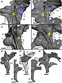 | 2,761 × 3,677 (2.93 MB) | FunkMonk | {{Information |description=Braincases of (a, c, e, f, h) Baryonyx walkeri (NHMUK PV R9951) and (b, d, g, i) Ceratosuchops inferodios (IWCMS 2014.95.1–3), in (a, b) right anterolateral and (c, d) right posterolateral, (e–g) right lateral and (h, i) posterior (right side) views, showing the major neurovascular features (and associated foramina) and braincase anatomy. Note that in Baryonyx, the separate cranial nerve trunks X—XI and XII open into a common fossa lateral to the occipital condyle,... |
File usage
The following pages on the English Wikipedia use this file (pages on other projects are not listed):