Jump to content

File:HeLa cell endocytic pathway labeled for EGFR and transferrin.jpg

Page contents not supported in other languages.
This is a file from the Wikimedia Commons
From Wikipedia, the free encyclopedia

Original file (3,259 × 3,510 pixels, file size: 7.17 MB, MIME type: image/jpeg)

Summary

Description
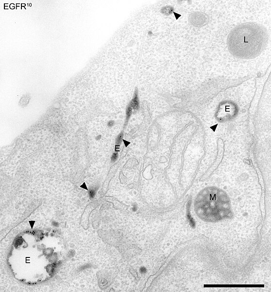
English: Compartments of the endocytic pathway in HeLa cells. Early endosomes (E), late endosomes/MVBs (M), and lysosomes (L) are visible. Epidermal growth factor receptors (EGFR) and transferrin (Tf) are labelled in the early endosomes.

Epidermal growth factor receptors are labelled with an antibody conjugated to 10 nm gold, applied to the cell surface. The cells were stimulated with EGF and allowed to internalize the receptors, bringing the gold with them. The image was taken 5 minutes after internalization, at which point most of the EGFR-gold has reached early endosomes (black dots, marked by arrowheads), but has not yet entered late endosomes or lysosomes. The cells are also loaded with transferrin conjugated to horseradish peroxidase (TfHRP). The HRP catalyses a reaction in the presence of DAB that produces a dark stain in the transferrin containing compartments in the image.

HeLa cells were preincubated for 1 h in serum-free medium. For the final 30 minutes the cells were incubated with TfHRP. The cells were then incubated with EGF and anti-EGFR 10 nm gold-conjugated antibody for 30 minutes at 4°C, washed, and incubated at 37°C for a further 5 minutes, all in the presence of TfHRP. Arrowheads; anti-EGFR 10 nm gold. Dark content; cross linked TfHRP. Bar, 500 nm.

Philips EM400 TEM

Methods for cell fixation and preparation for electron microscopy can be found in the reference below. Briefly, cells were room temperature fixed, a DAB reaction was done, and the cells were osmium stained, dehydrated and embedded en face in epon.

Related reference;

Doyotte, A., Russell, M.R.G., Hopkins, C.R., Woodman, P.G. (2005) Depletion of TSG101 forms a mammalian ‘‘Class E’’ compartment: a multicisternal early endosome with multiple sorting defects. J. Cell Sci, 118:3003-3017.
Date
Source Own work
Author Matthew R G Russell

Licensing

I, the copyright holder of this work, hereby publish it under the following licenses:
GNU head Permission is granted to copy, distribute and/or modify this document under the terms of the GNU Free Documentation License, Version 1.2 or any later version published by the Free Software Foundation; with no Invariant Sections, no Front-Cover Texts, and no Back-Cover Texts. A copy of the license is included in the section entitled GNU Free Documentation License.
w:en:Creative Commons
attribution
This file is licensed under the Creative Commons Attribution 3.0 Unported license.
You are free:
  • to share – to copy, distribute and transmit the work
  • to remix – to adapt the work
Under the following conditions:
  • attribution – You must give appropriate credit, provide a link to the license, and indicate if changes were made. You may do so in any reasonable manner, but not in any way that suggests the licensor endorses you or your use.
You may select the license of your choice.

Captions

Add a one-line explanation of what this file represents

Items portrayed in this file

depicts

1 September 2005

File history

Click on a date/time to view the file as it appeared at that time.

Date/TimeThumbnailDimensionsUserComment
current22:22, 3 November 2009Thumbnail for version as of 22:22, 3 November 20093,259 × 3,510 (7.17 MB)Putneybridgetube{{Information |Description={{en|1='''Compartments of the endocytic pathway in human HeLa cells.''' Early endosomes (E), late endosomes/MVBs (M), and lysosomes (L) are visible. Epidermal growth factor receptors (EGFR) and transferrin (Tf) are labelled in

The following page uses this file:

Global file usage

The following other wikis use this file:

Metadata